OUR PROJECTS
| Subproject 1 RNA Species and Networks for Diagnostics, Prognosis and Therapeutics |
|
|
Principal Investigator: Prof. Christoph Dieterich |
|
| Heart failure is a prevalent cause of death. Possible conditions that damage or overwork the heart muscle include coronary heart disease, diabetes, high blood pressure, cardiomyopathy and others. While these factors and combinations thereof weaken the heart, we do not understand why the heart fails at the molecular level and how we could best intervene in this process. Specifically, we do not know to what extent genetic and environmental contributions individually and jointly influence heart disease. We reason that the entire dynamic transcriptome would integrate and reflect these signals in an accessible way. Our first objective is to identify molecular disease states from human RNA sequencing data that correspond to a specific disease entity (e.g. dilative cardiomyopathy). We think of molecular disease states as transcript co-expression networks that respond in a coordinated way to environmental and disease conditions. As a first step, we need to compute personalized transcriptome representations using individual genome and transcriptome sequencing data. We need to control for known (e.g. age, gender) and hidden variation (e.g library preparation), which is unrelated to the phenotypic outcome. Additionally, deep sequencing empowers us to study RNA processing defects in parallel.In summary, we will arrive at a comprehensive characterization of protein-coding and non-coding RNA components, such as miRNAs, lncRNAs, μORFs and circular RNAs, many of which have been implicated in the context of heart disease. As a next step, we want to identify molecular subnetworks, which are deregulated in one or more studied heart disease settings. In other words, do different heart disease subtypes converge at, potentially novel, molecular nodal points, which could represent promising targets for therapeutic interventions? As a last step, we will investigate how these subnetworks could be utilized and targeted on the RNA level. Several studies describe the diagnostic, prognostic and possibly therapeutic values of ncRNAs in animal models. However, most of these outcomes have been the results of screening expeditions with little mechanistic insights and very few have been translated to the human System. It is our vision, to assist translation of previous and new findings to the clinics by using a quantitative systems-level approach on the RNA Level. 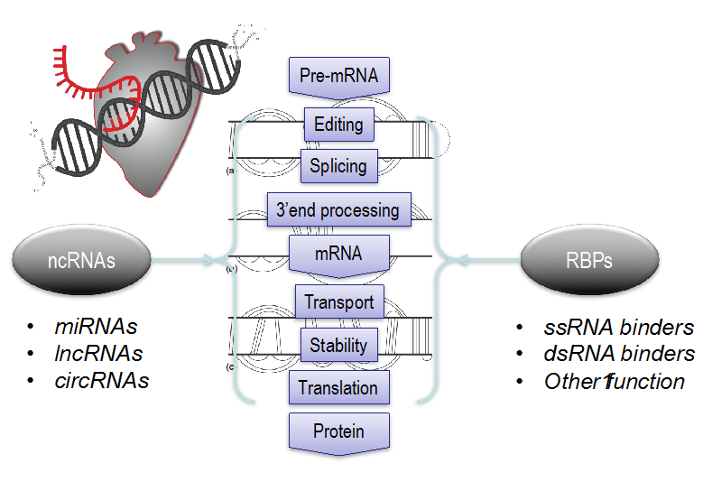 Figure 1 –The dynamic life of messenger RNA. Genetic and environmental perturbations are integrated at the RNA processing and network level, which is heavily regulated by non-coding RNAs (ncRNAs) and RNA-binding Proteins(RBPs). Figure 1 –The dynamic life of messenger RNA. Genetic and environmental perturbations are integrated at the RNA processing and network level, which is heavily regulated by non-coding RNAs (ncRNAs) and RNA-binding Proteins(RBPs). |
| Subproject 2 Structure-based Design of Peptide-based Pharmaceuticals against Striated Muscle Disorders |
|
|
Principal Investigators: Prof. Patrick Most, Prof. Rebecca Wade |
|
|
The objective of this joint research proposal is to leverage hybrid experimental and computer-aided methodologies for the structure-based modeling and development of peptide-based therapeutics against the fatal failure of striated muscle function in cardiomyopathies and skeletal muscular dys- trophies. To achieve this aim, we intend to use a novel biological mechanism by which the muscle- specific calcium sensor protein S100A1 improves both heart and skeletal muscle strength: S100A1 controls an intracellular protein network, in which downstream key members in turn regulate the contractile as well as the metabolic system (Pleger et al. 2013, Most et al. 2003, Most et al. 2006, Ritterhoff et al. 2015, Most et al. 2007, Prosser et al. 2010, Yamaguchi et al. 2011). Peptide do- mains located at the C-terminus of S100A1 seem to play an important role in calcium-dependent target binding. Our current understanding of how the intrinsically disordered S100A1 (Permyakov et al. 2011, Carvalho et al. 2013, Melville et al. 2017, Nowakowski et al. 2013, Nowakowski et al. 2011) may bind to and regulate its target proteins forms the basis for work package 1 (WP1): the systematic experimental analysis and mathematical modeling of the S100A1 protein-protein inter- action (PPI) network in heart and skeletal muscle to identify putative key targets and determine mutual binding sites. In work package 2 (WP2), iterative computational modeling will be employed to generate the three-dimensional molecular structures of S100A1/target complexes for structure- based identification of binding sites and design of ligands. Preliminary work along these lines has already produced a cell-permeable 20-mer peptide that is derived from S100A1’s disordered Helix 4-forming region (S100A1ct), which exerts biological and therapeutic activity in in vitro and in vivo disease models due to favourable physicochemical properties. As such, it serves as a starting point in work package 3 (WP3) for the structure-based design and refinement (Cardinale et al. 2011, Salo-Ahen et al. 2015, Panecka-Hofman et al. 2017, Stank et al. 2017, Martinez et al. 2015, Gdynia et al. 2016) of a portfolio of hit-to-lead optimized S100A1 peptide therapeutic(s) that sub- sequently will be tested in human iPSC-derived cardiomyocyte and skeletal muscle preparations from healthy and diseased individuals. Promising candidates will be subject to iterative cycles of experimental tests in molecular, cellular and animal disease models and refinement by computer- aided design to yield desirable pharmacokinetic, pharmacodynamic and toxicological properties. It is expected that peptide-based compounds resulting from this project will enter translational thera- peutic proof-of-concept studies in relevant large animal models of cardiomyopathies and muscular dystrophies and pave the way for early drug development. |
| Subproject 3 Research Data Warehouse (RWH) |
|
|
Principal Investigators: Dr. Nicolas Geis, Dipl.-Inform. Hauke Hund |
|
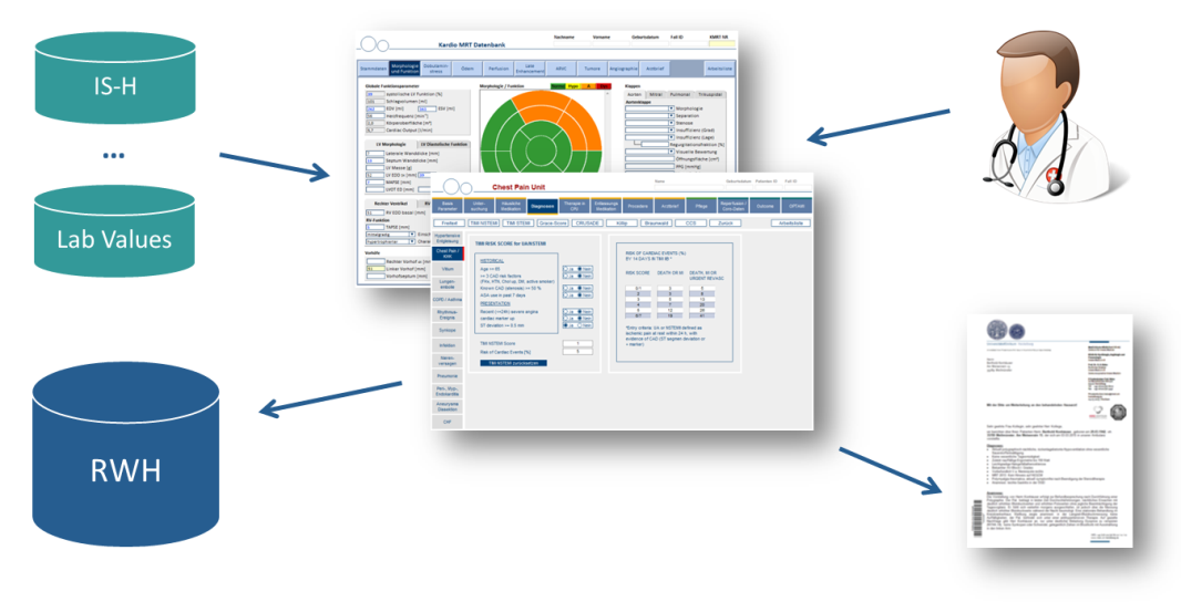
| To allow sophisticated clinical and translational research, clinical scientists need access to a multitude of information about patients’ characteristics such as diagnoses, genetic predisposition, disease phenotype, treatment, clinical course and outcome. To reduce the amount of time spent on reading written exams for the purpose of research, the “Research Data Warehouse” (RWH) was created in 2013 with the goal of providing a platform for analyzing structured patient data already existing in the department.
Figure 1: RWH architecture Existing processing pipeline (pseudonymization, extraction, transformation, loading (ETL)) for integrating various structured hospital data sources in the Research Data Warehouse. Pseudonymization is handled by the Trusted Third Party at Heilbronn University. So far, this platform has been used to perform retrospective studies as well as to select cohorts based on a specific phenotype for genetic analyses or for the enrollment in prospective studies.
Figure 2: Existing structured data entry systems at the Department of Cardiology This includes switching away from the digital type-writer approach which uses word processing software, to a routine use of information systems with structured data entry. Based on existing structured data entry systems developed for our Chest Pain Unit and Cardio-MRI/CT section, we propose to use this methodology also in other divisions such as ICH. (Institute for Cardiomyopathies Heidelberg), Cardio-Oncology, and others. These systems will enable semi-automatically generated written reports and simultaneously allow structured data collection for research purposes. |

| Subproject 4 Deep Learning-based Deformable Registration for Comparison of Magnetic Resonance Imaging Data |
|
| Principal Investigator: Prof. Sandy Engelhardt, Dr. Florian André Collaborating PIs: – |
|
|
Cardiac Magnetic Resonance (CMR) Images serve as rich sources of information about the patient’s disease state and is a widely used modality for diagnosis and disease monitoring. In clinical routine, analysis is mainly conducted quantitively, by extracting clinically relevant markers, such as volume estimates or ejection fraction, or qualitatively, by assessing scar tissue or the degree of fibrosis. However, there is a huge deficit of automatic and clinical deployable tools when images are to be compared to each other on a pixel-by-pixel level. For example, comparison is crucial for analysing follow-up images from the same patient to monitor longitudinal disease progression or to assess interventional outcome. Computational analysis that goes beyond the derivation of single one-dimensional parameters may be able to detect even subtle geometrical changes in these cases. Similarly, comparing an image against a population of patient images would allow for measuring geometric variability. In this case, a pathological case could be compared to a normal population to estimate the difference in form of a vector field. Apart from that, the geometric variability within a population could be more appropriately assessed.
|

| Subproject 5 Population-scale Predictive Networks of Myocardial Dysfunction and Heart Failure |
|
|
Principal Investigator: Prof. Benjamin Meder |
|
| Genetic variations have a fundamental impact on phenotypic variability. In cardiovascular disease, especially in HF and cardiomyopathy, not only single high penetrance mutations result in morbidity and mortality, but also highly frequent genetic background variations can modify cardiac structural and functional parameters and hence affect disease outbreak and Progression. As an example may serve the elastic giant protein Titin. Truncating and non-truncating variants are highly frequent in dilated cardiomyopathy (DCM; TTN-TV frequency: 14-20%), but also in normal populations (1-3%), theoretically rendering up to 100 Million persons in the European Union prone to develop heart failure due to genetic mechanisms. Importantly, the genetic variant seems not to be sufficient to provoke clinically manifest disease by itself, but can result in undetected cardiac limitations or HF outbreak due to different and with age increasing noxae. With the revolutionary advances in Omics technologies and computational approaches, cardiovascular research gains new opportunities in finding key mechanisms for heart failure, enabling identification of novel diagnostic and therapeutic targets. To better understand the nodal players that drive heart failure, it is reasonable to investigate affected patients but also normal controls on a population scale. The role of the proposed project is to combine data from 1) clinical phenotyping, 2) large-scale population studies (Big Data), 3) functional cardiovascular imaging and 4) omicsdatasets to generate multi-scale mathematical models as predictors of the impact of distinct genetic and epigenetic variants on structural and functional sarcomeric components (Molecular Modeling) and subclinical/clinical phenotypes (Disease Modeling). In this project, we will systematically combine clinical and big data on a population-scale, molecular modeling of genetic mechanism and associated molecular networks and disease modeling based on personalized computer equations of individuals characterized on the different levels. By this strategy, it is envisaged to advance the current knowledge about genetic and phenotypic variability and identify individuals at exaggerated risk for development of HF. This would have great Impact not only for cardiology, but many other disciplines including oncology (provoking HF due to chemotherapy) or gynecology (HF in the course of pregnancy). 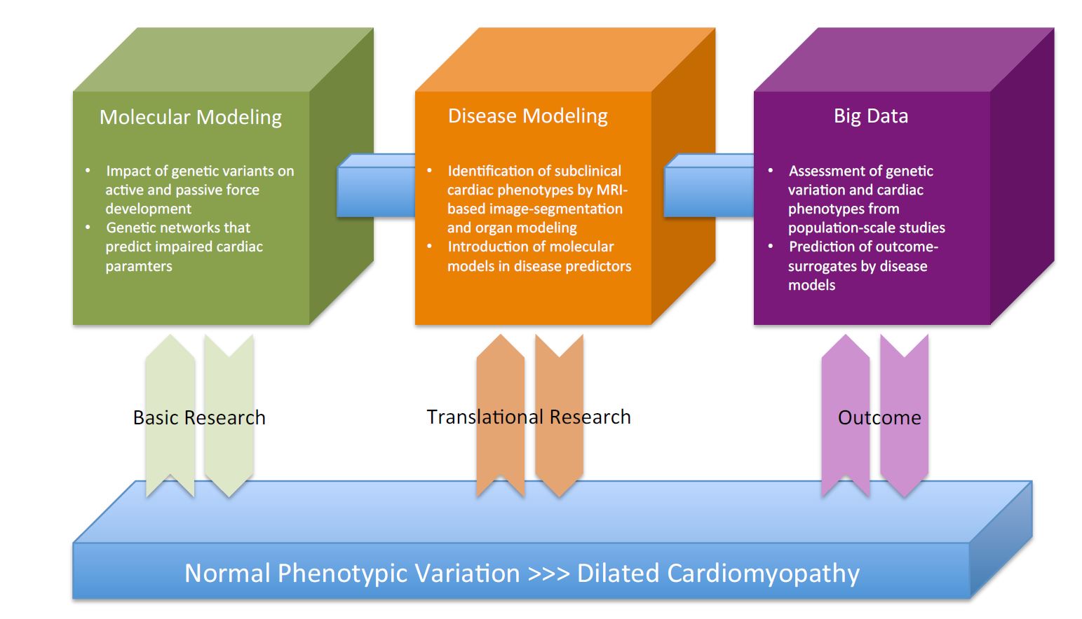 Figure 1 – Strategy to dissect the impact and predictive power of multi-scale computational molecular networks on clinical penotypes from individual patients to the population scale. Figure 1 – Strategy to dissect the impact and predictive power of multi-scale computational molecular networks on clinical penotypes from individual patients to the population scale. Figure 2 – Datasets illustrating the concept of multi-omics in translational cardiovascular research (based on Meder et al. 2017) Until now, phenotypic data were used to generate computational models of heart failure, which we will expand by using molecular data for cardiomyocyte simulation. Figure 2 – Datasets illustrating the concept of multi-omics in translational cardiovascular research (based on Meder et al. 2017) Until now, phenotypic data were used to generate computational models of heart failure, which we will expand by using molecular data for cardiomyocyte simulation. |
| Subproject 6 Optimization and visualization support system for cardiac arrhythmia management |
|
| Principal Investigators: Dr. Ann-Kathrin Rahm Collaborating PI: Prof. R. Herzog |
|
|
The combination of an ageing population due to the demographic change together with ongoing medical progress in the treatment of advanced HF continuously increases the number of patients that are prone to cardiac arrhythmias. This development poses an urgent need for optimized diagnostic and therapeutic approaches. Based on the previous work, the aim of this subproject is to continue the use of already developed mathematical models and apply them to clinical use.
|

| Subproject 7 Cognition and Uncertainty Quantification for Numerical Heart Simulation |
|
| Principal Investigator: Prof. Vincent Heuveline Collaborating PIs: Prof. R. De Simone, Prof. S. Engelhardt, Prof. C. Rother |
|
|
Over the last two decades, medicine and surgery were characterized by a considerable number of newly developed techniques and supportive instruments in an effort to assist, quantify and improve processes in the medical, clinical and interventional context. In interventional cardiology, for instance, percutaneous, valvular and vascular devices today enable increasingly effective minimally invasive treatment, avoiding surgery. Simultaneously, medical imaging and segmentation techniques allow surgeons to obtain a clearer understanding of the organ’s morphology that they operate on. Yet, functional analysis is mostly up to the experience of the physician. Particularly with the mitral and tricuspid valve, all modifying techniques are highly challenging with regard to procedure choice and planning. Thus, a simulation of therapeutic options for individual patients could further improve efficacy and outcome and reduce complications. Functional mathematical modeling and simulation, allows to virtually reproduce and predict the behaviour and function of organs. As an example, soft tissue simulation may give insights into the biomechanical behaviour of the mitral and tricuspid valve during or after surgical or interventional manipulation. Similarly, computational fluid dynamics or fluid structure interaction simulation allows to depict flow processes in the cardiovascular system. In a more complex virtual pharmacology simulation, the effect of hemodynamically active substances in various decompensated valvular diseases could be predicted. Such obtained simulation-based information is, however, only useful and usable if the underlying simulation scenarios are patient-specific and cognition-based, and if the uncertainty in the simulated results is quantifiable. Therefore, it is important that the large amounts of medical patient data is properly analyzed and processed through dedicated data mining and information processing Workflows. Simulation scenarios not only need to be comprehensive in terms of the available patient data and holistic under the consideration of medical knowledge. They can also be patient-individually calibrated, e.g., through methods for Data Assimilation. Moreover, by means of a semantic, cognitive-technical description of surgery and intervention modeling and simulation workflows, patient data can be assigned and processed intelligently. Knowing that both the medical data and the numerical simulation are subjected to measurement inaccuracies and uncertainties, techniques for uncertainty quantification (UQ) – both for describing and quantifying noise in the data as well as for assessing model simplifications and simulation inaccuracies – have to become an essential component part of any medical simulation system in order to obtain reliable simulation results. Lastly, in order for an adequate applicability and usability of simulation-enhanced surgery assistance systems, biomechanical simulations need to be fully integrated into the clinical treatment workflow and into traditional surgery assistance systems. They then have the potential to not only drive forward a more comprehensive and holistic medical analysis and diagnosis, but also to significantly support and improve operative surgery planning and intervention. The aim of this project is to develop new methods and high performance computing software components for cognition-guided surgery simulation with uncertainty quantification.
|

| Subproject 8 Comorbidities in Heartfailure – A Network Approach |
|
|
Principal Investigator: Prof. Julio Saez-Rodriguez, PD Dr. Jobst-Hendrik Schultz |
|
| This interdisciplinary subproject aims to develop a co-morditity-based understanding based on cases of heart failure (HF) in a real-life clinical and outpatient departement. Data derived from departments of internal medicine of the University Hospital Heidelberg will be analyzed with the aim to construct co-morbidity-associated disease models that can be incorporated into a personalized systems medicine approach. In cooperation with the Research Ware House (RWH, SP 3) and data sources from the clinical-wide hospital information system, which partially relies on human computer interaction, patients suffering systolic or diastolic HF will be retrieved according to the state-of-the-art definitions. A network approach then investigates and evaluates a patient-specific co-morbidity-/risk profile with clinical or subclinical impact on cardiac function based on a six-dimensional model of patient characteristics that comprises environmental , psycho-social (e.g. depression), neuro-vegetative (e.g. HRV, cortisol), inflammatory (e.g. CrP), somatic (e.g. weight), and organ-specific (e.g. nephrological, endocrine, etc.) factors. Molecular data will be analyzed with functional genomics methods. Machine learning, supported by crowdsourcing in the form of a DREAM challenge will be utilized to predict factors affecting HF. The network will comply with high standards of medical data protection. The applicability of this interdisciplinary approach will be evaluated with a clinical study conducted in different units of the university hospital. |
| Subproject 10 Multiparametric single-cell morphological analysis of cardiomyocytes for translational cardiovascular research |
|
| Principal Investigators: PD Dr. Matthias Konstandin, Prof. Stefania Petra, Dr. Kai Polsterer Collaborating PIs: — |
|
|
Heart failure is the endpoint of a wide range of cardiac diseases. It is a complex clinical syndrome that involves cellular as well as metabolic and neurohormonal communication. To prevent disease progression, it is important to diagnose it early and start a therapy that is specific to the patient’s aetiology. In the present project we aim to complement the patients diagnostic workup by the analysis of morphological features of cardiomyocytes treated in vitro with the patient‘s plasma to gain prognostic or diagnostic value. Our intent is to apply machine learning and mathematical imaging techniques in order to fully exploit the information present in this morphology. Our starting point is C-MORE, a complex cellular morphology-based assay on lab cultured neonatal rat cardiomyocyte (NRCMs). We adopt C-MORE as a baseline to help us identify pathomechanisms in cardiomyocytes that are incubated with patient blood plasma and/or pharmacological substances mimicking neurohumoral activation in vitro. The current workflow will be enhanced with state-of-the-art machine learning methods. Currently used features will be complemented by features automatically learnt by convolutional neural networks (CNNs). Statistical classification and context-sensitive image segmentation, based on these features, will improve the transparency of the automated decision-making process. Our goal is to develop a diagnostic tool with an “explainable AI” component at its heart.
|
| Subproject 11 Machine learning-based interpretation of complex genomic traits |
|
| Principal Investigators: Prof. Oliver Stegle, Dr. Jan Haas Collaborating PIs: — |
|
|
Coming soon |